
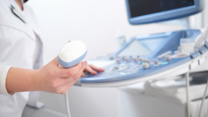
Ultrasound imaging, also known as sonography, is a versatile and non-invasive diagnostic tool widely used in healthcare to visualise internal body structures. It employs high-frequency sound waves to create images of the inside of the body, aiding in the diagnosis, treatment, and monitoring of medical conditions.
Principles of Ultrasound
Ultrasound works by sending sound waves into the body using a transducer. These sound waves bounce off tissues, organs, and fluids at varying speeds, returning echoes to the transducer. The ultrasound machine then interprets these echoes into real-time images. Since the method doesn’t involve radiation, it’s particularly favoured for examining developing foetuses in pregnant women.
Applications in Medicine
- Obstetrics and Gynaecology: Ultrasound is perhaps best known for its role in obstetrics where it’s used for checking the health of a developing foetus. It helps in assessing foetal growth, determining the due date, identifying multiple pregnancies, and diagnosing potential complications. In gynaecology, it’s used to examine the uterus, ovaries, and fallopian tubes, aiding in the diagnosis of conditions like fibroids, cysts, and ectopic pregnancies.
- Cardiology: In cardiology, echocardiograms utilise ultrasound to create images of the heart, revealing information about its structure, function, and blood flow. This is crucial for diagnosing heart conditions such as valve problems, cardiomyopathy, and congenital heart disease.
- Abdominal Exams: Ultrasound is used to examine internal organs like the liver, kidneys, gallbladder, spleen, and pancreas. It helps to diagnose stones, infections, tumours, and other abdominal ailments.
- Musculoskeletal (MSK) Ultrasound: This type of ultrasound assesses muscles, tendons, ligaments, and joints. It’s increasingly used for diagnosing tears, sprains, and other injuries, and ultrasound guided cortisone injections in pain management.
- Vascular Ultrasound: Vascular sonography helps to visualise the body’s veins and arteries. It’s instrumental in identifying blockages (stenosis) and abnormalities like aneurysms or thromboses, and in evaluating the effectiveness of vascular procedures.
Advantages of Ultrasound
Ultrasound holds several advantages over other diagnostic methods:
- Safety: Ultrasound doesn’t use ionising radiation, making it safer than X-rays and CT scans, especially for pregnant women and those requiring multiple examinations. This characteristic significantly reduces the risk of radiation-induced complications and makes ultrasound a preferred choice for monitoring ongoing conditions and foetal development throughout pregnancy.
- Real-time Imaging: Ultrasound provides real-time imaging, making it extremely valuable for guiding minimally invasive procedures such as biopsies. This capability allows healthcare providers to observe the movement of instruments in real time, adjust their techniques on the fly, and ensure the accuracy and safety of the procedure, thereby improving patient outcomes and reducing the risk of complications.
- Portability: Modern ultrasound machines can be portable, allowing for bedside examinations and use in remote areas where traditional imaging facilities may be inaccessible. This portability is crucial in emergency and rural medicine, enabling rapid, on-the-spot diagnostic assessments and medical decision-making, which can be lifesaving in critical care situations.
Limitations
Despite its numerous benefits, ultrasound has limitations. It can be hindered by the presence of gas or fat, as these can obscure the sound waves, making it difficult to obtain clear images. Also, the quality of the images depends significantly on the operator’s skill.
In summary, ultrasound is a fundamental tool in modern healthcare, vital for its diagnostic precision, safety, and versatility. It continues to evolve with advancements in technology, enhancing its capabilities and applications in medicine.
